


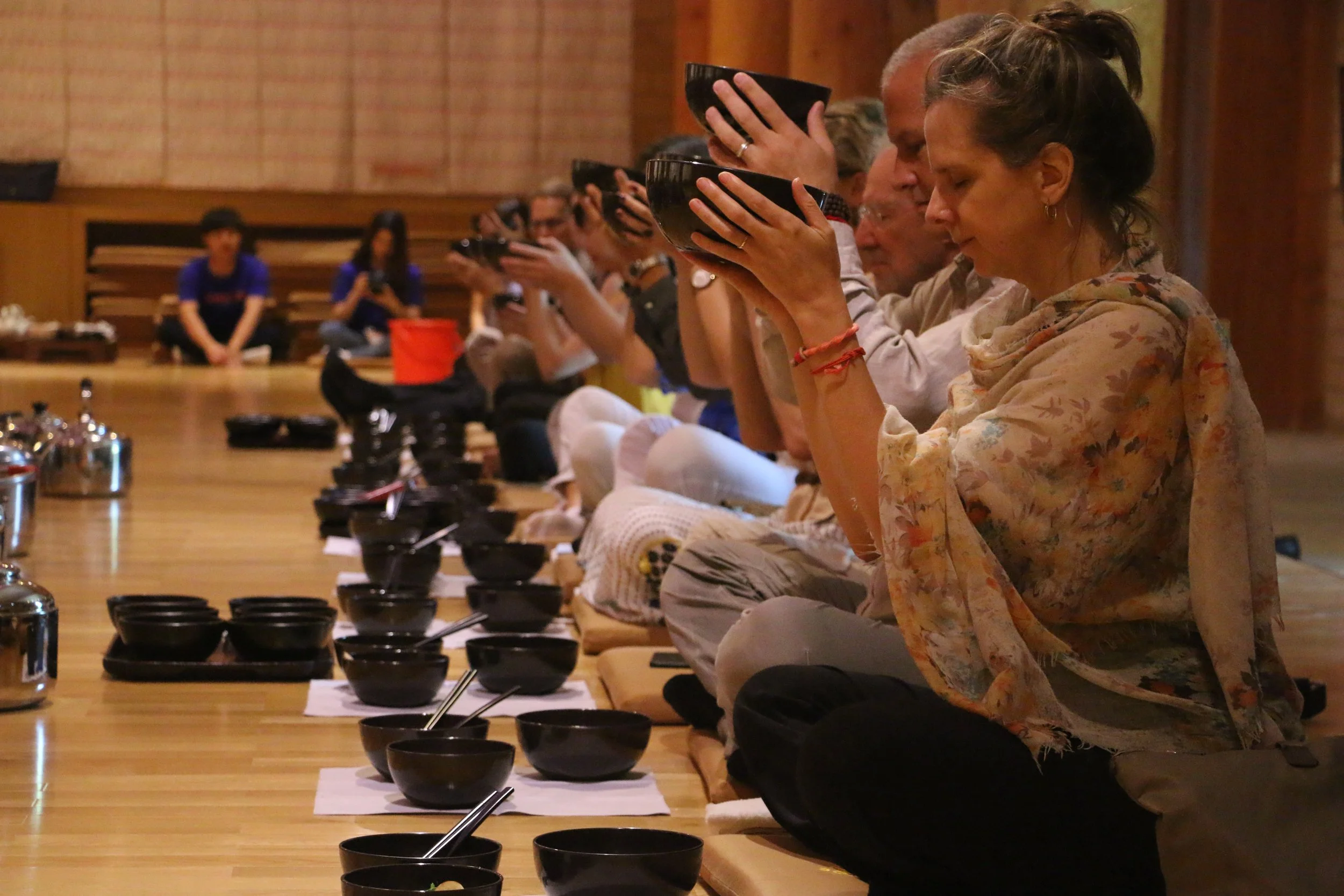


Religion, as humanity’s oldest expression of values, identity and community, has always been a mediated practice. In contemporary society religious ideas are communicated, learned, represented, enacted and resisted through various forms of media. Religion circulates through social media, is discussed in the news and becomes a source of imagery for film and television.
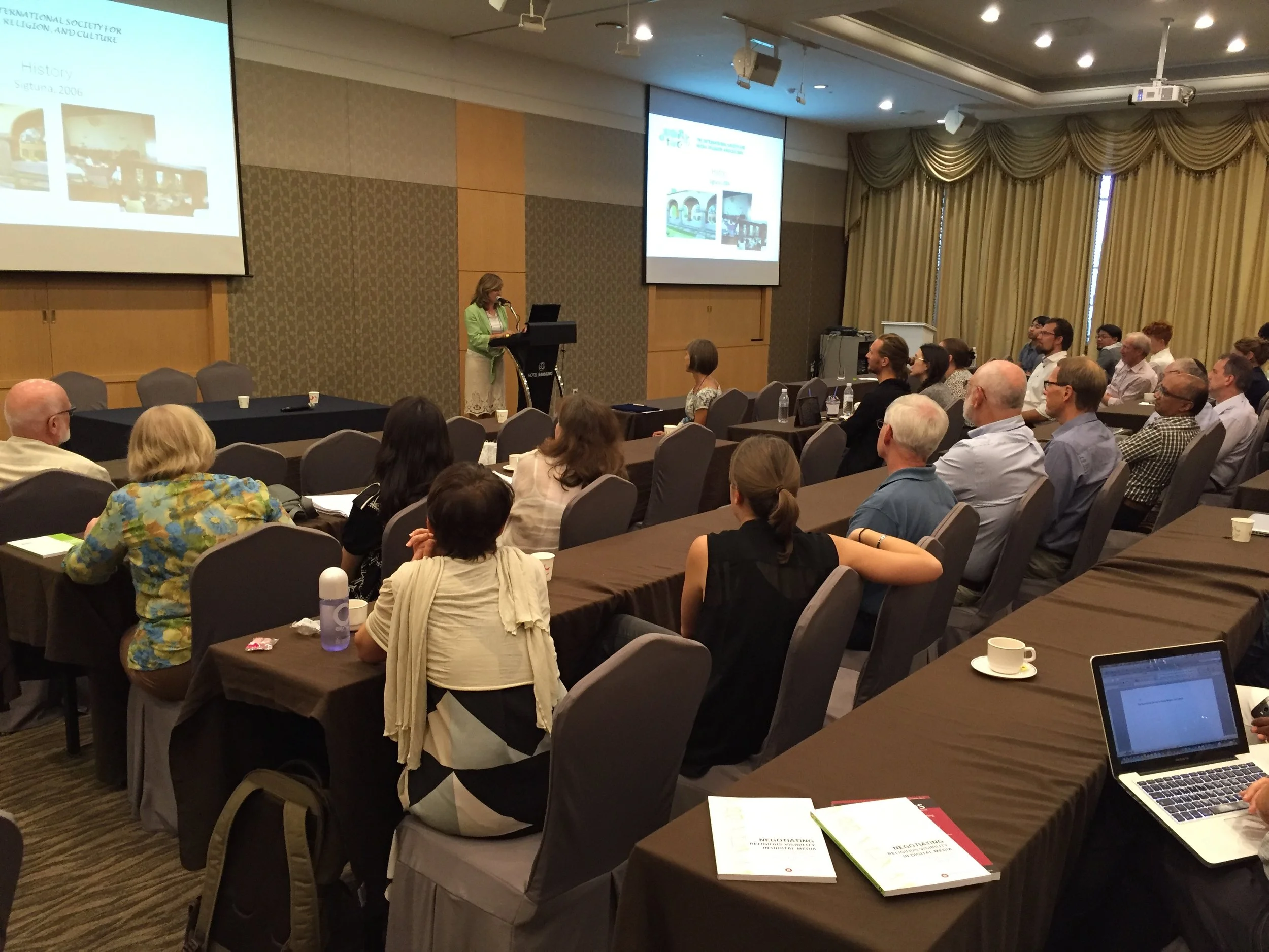
The International Society for Media, Religion and Culture (ISMRC) was founded in 2010 out of a series of conferences on the interplay between media, religion and culture starting in 1994. Today the ISMRC is a vibrant research community of scholars with a large variety of international, disciplinary and religious backgrounds, covering topics such as journalism on religion; religion, popular culture and entertainment; and digital religion.
The ISMRC organizes biennial conferences and the Journal of Religion, Media and Digital Culture (RMDC) is published by Brill in cooperation with the ISMRC.
-

About the Society
Current board members
Journal of Media, Religion and Digital Culture
-

Conferences
Past and future conferences
-

Membership
Benefts of being a member
-

Community
Member news and updates, newsletter, recent publications
-

Oren Golan
University of Haifa, Israel
-

Katja Valaskivi
University of Helsinki, Finland
-

Kristin Peterson
Boston College, USA
-

Marta Kołodziejska
University of Warsaw, Poland
-

Deborah Whitehead
University of Colorado Boulder, USA
-

Alessandra Vitullo
Sapienza University of Rome, Italy
-

Rebekka Rieser
University of Zurich, Switzerland
-

Feeza Vasudeva
University of Helsinki, Finland
-

Sana Patel
Rice University, USA
-

Miriam Diez Bosch
Ramon Llull University, Spain